Determine Which of the Following Is the Oculomotor Nerve's Function.
Determine if the patient can see anything. Somatosensory information touch pain from the face and head.

Learn About The Spinal Cord And Its Anatomy And Parts Find Out About The Spinal Cord Function What It Does And Where Do Spinal Cord Spinal Cord Anatomy Spinal
They bring sensations from proprioceptors in the eye muscles.

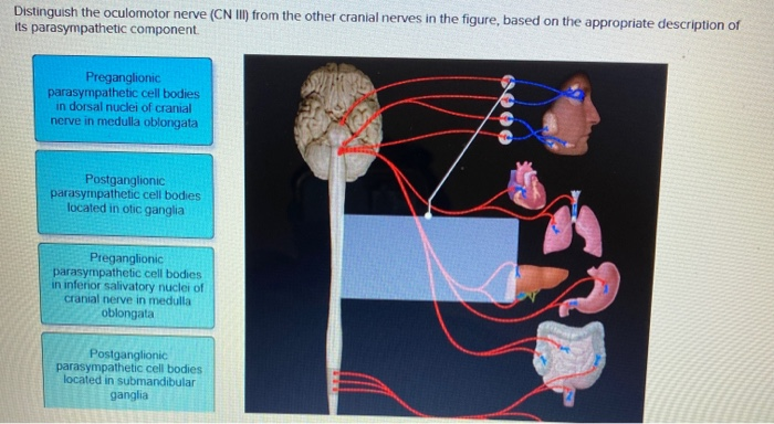
. These will be included as the culminating activities of the lab on cranial nerves. Third Fourth sixth Nerve Test. The visceral motor function provides parasympathetic innervations to the constrictor pupillae and ciliary muscles.
Have the patient distinguish between green and red colors. The oculomotor nerve or the third cranial nerve is associated with eye motor functions4. Have the patient distinguish between green and red colors B.
Mydriasis as the parasympathetic fibres of the oculomotor nerve surround the motor fibres. The oculomotor nerve is entirely motor It is responsible for lifting the upper eyelid. Select which cranial nerve has sensory fibers that monitor blood pressure at the carotid Sinus.
The 3rd cranial nerves are pure motor nerves. Determine if the patient still has night vision. Which of the following would help to determine if the oculomotor nerve was damaged.
It is called common ocular motor nerve and sends orders to the muscles involved in eye movement causing the pupil to dilate or contract. The abducens nerves A transmit pain impulses from the teeth. Determine if the patient can see anything C.
It is a motor nerve that sends signals from the brain to the muscles. The oculomotor nerve is responsible for the majority of eye and eyelid movements although the trochlear nerve and abducens nerve also contribute to eye movements. To determine if there were any improvements in student learning I included the same questions on cranial nerves on the exam this year attached.
This nerve innervates to the upper eyelid somatic the pupil and lens parasympathetic and the eye muscles for gaze fixation and visual tracking somatic5. Any damage to this nerve results in problems related to sight and vision. Simply from the name then it is easy to know that the oculomotor nerve will innervate muscles that move the eye itself or components of the eye.
Oculomotor nerve or cranial nerve III. It is part of the autonomic nervous system which supplies innervates many of your organs including the eyes. If the oculomotor nerve is compressed which symptom is the first to be seen and why.
Optic nerve It carries visual information from your retina to your brain. Oculomotor nerve It controls most of your eye movements along with the way your pupil constricts and the ability to keep your eyelid open. Anatomy and Physiology questions and answers.
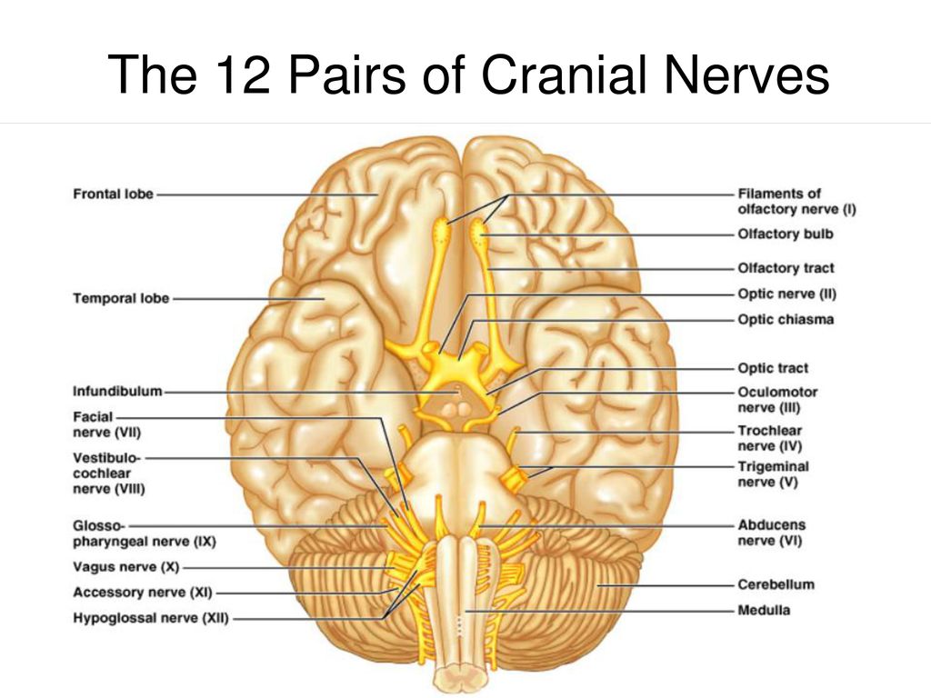
The oculomotor nerve originates from the oculomotor nucleus and accessory parasympathetic nucleus in the midbrain6. The 12 cranial nerves and their functions are. Olfactory nerve It controls your sense of smell.
Optic nerve or cranial nerve II. Test 4 Brain Cranial Nerves - AP 1. Pupillary contraction Determine which of the following is the oculomotor nerves function.
Why is the eye in a down and out position in oculomotor nerve palsy. They are Lower Motor Neurons LMN second order neurons. The optic nerve II is the agent of vision.
Asked Sep 27 2016 in Anatomy Physiology by polishaddict. The oculomotor nerve is the third cranial nerve CN III and one instance in which the name is a clear indication of the function of the nerve Oculo pertaining to the eye motor producing movement. Lateral rectus abducens is still intact and so the pulls the eye out and the superior oblique.
Have the patient cry E. This transmits visual information from the eyes to the brain and vice versa. They sit at the level of the tentorium.
The oculomotor nerve helps control muscle movements of the eyes. They control ocular movements so considered together. Cranial Nerve Function and the Cranial Nerve Flowchart will be required assignments.
Which of the following would help to determine if the oculomotor nerve was damaged. Move the eye inferiorly and laterally Conduct impulses for vision Pupillary constriction Abduct the eye Reset Zoom. This nerve is the fourth set of cranial nerves CN IV or cranial nerve 4.
Ptosis drooping of the upper eyelid of the left eye would be caused by damage to the A facial nerve. Asked Nov 9 2016 in Health Professions by Chelsea. It is an afferent fiber and transmits information to the brain from the eye.
They are mixed nerves. E determine if the patient still has night vision Answer. Abducent nerve innervates.
Oculomotor nerve helps in the movement of the eye. They control eye muscles on the same side of the body ipsilateral. CN IV works with the oculomotor nerve and other eye muscles to control eye movement.
Number Name Function Location. The oculomotor nerve has two distinct functions the somatic motor that supplies four of the six extraocular muscles of the eye and the levator palpabrae superioris muscle of the upper eyelid. Determine which of the following is the oculomotor nerves function.
When inspecting a clients eyes the nurse assesses the presence of cranial nerve III oculomotor cranial nerve by observing as the eyelids open and close bilaterally and also by testing. Oculomotor nerves are responsible for which of the following functions. The oculomotor nerve controls eye movements.
Have the patient look superiorly and inferiorly D. The two 3rd cranial nerves oculomotor nerves are located at the top of the brainstem - one to the right and one to the left. It is the shortest of all pairs.
It innervates the ipsilateral dorsal ventral and medial recti muscls and the ventral oblique muscle. Damage to this nerve leads to distortion in vision or double vision and even problem in the coordination of eyes. A 13-year-old child exhibited retarded growth reduced metabolism lack of normalreproductive gland development inability to regulate water intake or water elimination from the body and an uncontrolled appetite.
Fibers of these nerves take origin from a series of nuclei which begin in the floor of sylvian aqueduct and extending up to the fourth ventricle. What is the function of the oculomotor nerve. The oculomotor nerve provides movement to most of the muscles that move the eyeball and upper eyelid known as extraocular muscles.

Ot 716 Cranial Nerves Unit 3 Flashcards Quizlet

Cranial Nerves Quizzes And Labeling Exercises Cranial Nerves Facial Nerve Nerve Anatomy

Medical Encyclopedia Structure And Function Brain Spinal Cord And Nerves Caudate Nucleus Spinal Cord Structure And Function

Cranial Nerves Nerve Brain Anatomy

Examining The Trigeminal Nerve Medical School Studying Nursing School Survival Nursing School Studying

Sheep Brain External Anatomy Ventral Dollar Store Christmas Crafts Abducens Nerve Hypoglossal Nerve
![]()
Oculomotor Nerve Cn Iii Anatomy Function And Pathway Kenhub
Solved Distinguish The Oculomotor Nerve Cn Iii From The Chegg Com
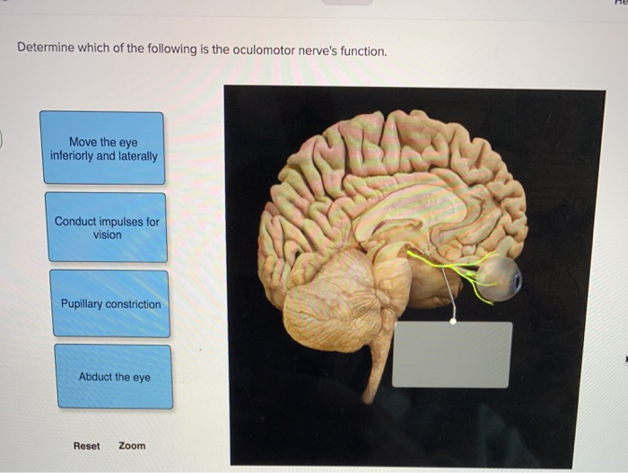
Solved Determine Which Of The Following Is The Oculomotor Chegg Com
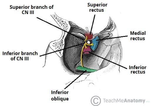
The Oculomotor Nerve Cn Iii Course Motor Teachmeanatomy

Dear Nurses Simplifying The Cranial Nerves Cranial Nerves Craniosacral Therapy Nursing School Tips

Functions Of The Cerebral Cortex Cerebral Cortex How To Memorize Things Powerful Computer
(179).jpg)
Quiz On Cranial Nerves Mcq Trivia Proprofs Quiz

Cranial Nerves Tricks Mnemonics Pares Craneales Anatomia Del Esqueleto Humano Anatomia Medica

Cranial Nerves On Models Labeled Brain Model Somso Nervous System Anatomy Anatomy And Physiology Brain Anatomy
Omni Body Healthcare Cn Iii The Oculomotor Nerve The Oculomotor Nerves Third Cranial Nerves Originate In The Midbrain And Eventually Supply The Pupil Constrictor Eyelid Adductor

Ventricles Of The Brain Anatomy Model Function And Mnemonic Brain Anatomy Brain Facts Anatomy
Cranial Nerves Anatomy Function Olfactory Optic Oculomotor Trochlear Trigeminal Abducent Science Online

Cranial Nerves Brain Anatomy Human Anatomy And Physiology Medical Anatomy


Comments
Post a Comment